
Transvaginal ultrasound: how it works and why it is important
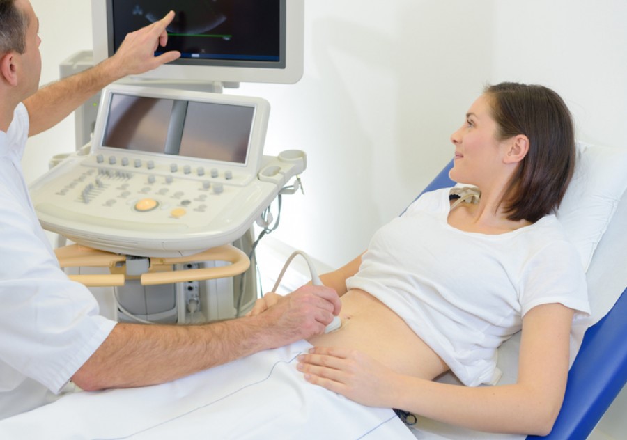
Transvaginal ultrasound is a very common non-invasive gynaecological diagnostic test. It can be two- or three-dimensional, is usually associated with a gynaecological examination and enables the internal reproductive organs and surrounding organs to be assessed using a probe positioned in the vagina
Originally used to detect malformations of the uterus, it is now essential for detecting and diagnosing a variety of disorders and diseases.
Transvaginal ultrasound: an essential examination
Transvaginal ultrasound is a particularly useful examination as it enables the uterus, ovaries and fallopian tubes (or salpingi), as well as the surrounding organs such as the bladder, ureters, rectum and sigma, to be observed safely and quickly.
This is why gynaecological ultrasound examinations now have many indications in daily practice:
- in the diagnosis of neoformations or malformations suspected on an anamnestic and objective basis in the genital organs;
- in the diagnosis of women suffering from chronic pelvic pain or dysfunctional pathologies (irregular menstrual cycles) or infectious processes affecting the internal genitals;
- in the differential diagnosis with other abdomino-pelvic pathologies in urgent conditions (appendicitis, diverticulitis, colitis);
- in the diagnostic pathway of peri- and post-menopausal patients with atypical bleeding, helping to determine the characteristics of the endometrium and the uterine cavity (suspicion of endometrial neoplasms or search for polyps);
- in the surveillance of the ovaries and endometrium in women with a genetic predisposition to ovarian carcinoma or family syndromes for tumours (BRCA mutated and/or Lynch syndrome);
- in the diagnostic pathways of infertile patients (diagnosis, monitoring and assistance in assisted reproduction techniques);
- in the monitoring of medical therapies (tamoxifen or hormonal therapies), in the control of surgical outcomes (after myomectomies) and in the diagnostic pathways of patients suffering from pelvic static disorders (urinary incontinence, severe constipation).
How transvaginal ultrasound is performed
Transvaginal ultrasound is based on the action of sound waves and is neither dangerous nor painful for the patient.
In fact, the examination is usually carried out at the same time as the gynaecological examination (unless otherwise instructed by the specialist), using a probe capable of emitting high frequency ultrasound inserted into the patient’s vagina, lying on the couch in the gynaecological position.
The sound waves emitted by the probe are reflected by our internal organs and processed as images on the ultrasound monitor, allowing the specialist to assess the condition of the areas of interest.
Transvaginal ultrasound can be carried out at any time, even when menstrual flow is present, on patients who have already started sexual activity; it cannot be performed if the patient has never had sexual intercourse.
In this case, it is preferable to subject the patient to transabdominal ultrasound with a full bladder, which still allows good observation of the same pathologies.

But can it be dangerous to perform transvaginal ultrasound during pregnancy?
Absolutely not: transvaginal ultrasound is commonly used in the first few weeks of pregnancy to observe the embryo and correctly date the pregnancy (while transabdominal ultrasound offers better images later on) and to carry out cervicometry, i.e. measuring the length of the cervix, which is essential for monitoring the risk of miscarriage in the first trimester of pregnancy.
In cases of blood loss it is used to assess a risk of miscarriage or unfortunately an ongoing miscarriage.
First and second level transvaginal ultrasound: what are the differences?
Transvaginal ultrasound can be either level 1 or level 2.
First level transvaginal ultrasound is performed at the same time as the gynaecological examination and is used to detect the presence of any conditions or diseases.
It may be necessary to follow this up with a level 2 transvaginal ultrasound scan, which provides the specialist with more precise information on the element in question.
The second level ultrasound is therefore requested as an in-depth examination following the gynaecological examination and the first level ultrasound, when there is a suspicion of a pathology that requires further checks and it is carried out with different timing and procedures.
Read Also:
Pre-Hospital Ultrasound Assessment In Emergencies
DVT Ultrasound Fails Too – Is It Enough To Detect Real Disease?