
Tumors of the nervous system: craniopharyngioma
Craniopharyngioma is a rare tumor that arises in a region rich in nerves and blood vessels
Craniopharyngioma is a neoplasm arising from the embryonic remnants of the primitive intestine (Ratke’s pouch)
They may persist after birth at the level of the tissues surrounding the sella turcica region (bony formation located inside the skull with a cavity that houses the pituitary gland).
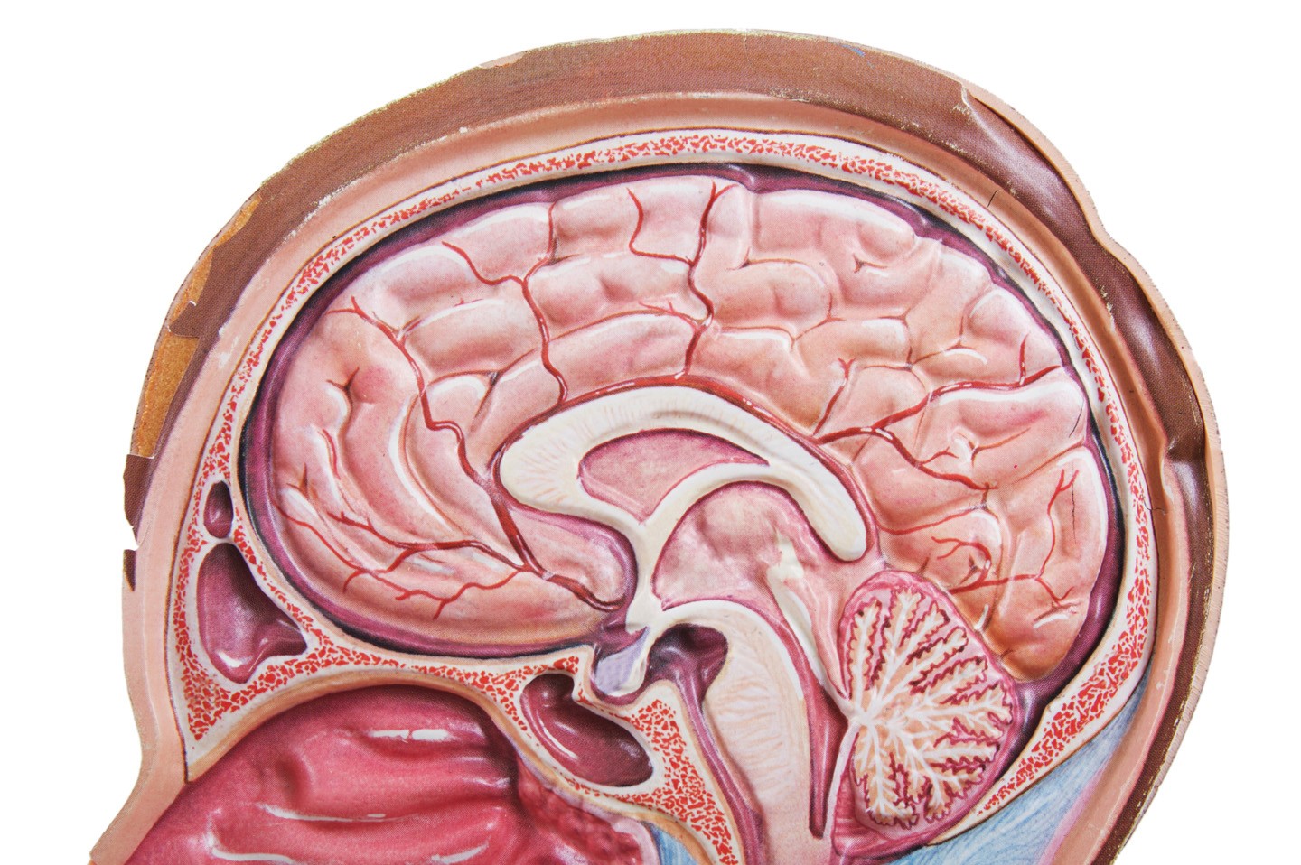
The saddle and parasellar region contain numerous vital nerve and vascular structures: the arteries of the circle of Willis (which distribute arterial blood to the entire brain), the optic nerves (which conduct visual information), the pituitary gland and its pedicle (which regulate the production of the body’s hormones), and the hypothalamus (the center of integration of brain functions related to behavior, regulation of metabolism, and other vital functions).
Although craniopharyngioma has a low growth rate, the fact that it arises in contact with anatomical structures of great functional importance makes it extremely dangerous.
It is a rather rare neoplasm, accounting for about 5% of central nervous system neoplasms in children.
The symptoms of craniopharyngioma depend on compression of nerve structures around the sella turcica
Generally, reduced vision, diabetes insipidus (production of high volumes of dilute urine), changes in body weight (usually a tendency toward obesity), fatigue, and puberty alterations (due to hormonal changes) may occur.
Some children have a hydrocephalus condition with typical symptoms (headache, vomiting, decreased responsiveness).
Diagnosis is made by Computed Tomography and Magnetic Resonance Imaging, which allow detailed identification of the characteristics of the neoplasm and the relationship to nerve and vascular structures.
Blood and urine tests allow assessment of hypothalamic-pituitary hormone production.
CHILD HEALTH: LEARN MORE ABOUT MEDICHILD BY VISITING THE BOOTH IN EMERGENCY EXPO
There are two histologic variants of craniopharyngioma:
- Papillary: typical of adulthood.
- Adamantinomatous: typical of pediatric age, characterized by a tendency to infiltrate nerve and vascular structures, with frequent presence of calcifications and cystic components. Sometimes the cystic components (filled with a brownish oily fluid very rich in protein) are voluminous and are responsible for the symptoms.
The treatment of craniopharyngioma is multidisciplinary in nature and involves from the earliest stages of diagnosis a confrontation between various specialists
Complete surgical removal, when feasible, is a key step in the care of these children.
Surgical maneuvers, however, must always preserve neurological and endocrine function as much as possible, especially in children.
There are no effective drugs to combat pediatric craniopharyngiomas, and therefore, when surgery is not indicated, radiation therapy is used to help control the disease.
A special form of radiotherapy performed with protons instead of photons is currently the treatment of choice for craniopharyngiomas that cannot be completely removed because it is burdened with fewer long-term side effects than traditional radiotherapy.
At present, there is no shared international treatment protocol, but the treatment philosophy is to stabilize the disease while minimizing clinical complications (loss of vision and development of obesity or other endocrinological problems).
In selected cases, classic microsurgery may be combined with transnasal endoscopic procedures
With regard to cystic components, there is recent evidence of the usefulness of proceeding to their emptying by placement with a stereotactic apparatus (a surgical tool that allows surgery inside the skull) of intracystic catheters and local administration of interferon, a substance capable of blocking the production of cystic fluid.
Hormonal defects caused by the disease can be adequately corrected with specific drugs.

Recent molecular studies have identified the presence of a specific mutation (BRAFV600E) in papillary craniopharyngiomas.
Targeted therapies can be considered in this subgroup of patients.
In the adamantinomatous form, which is typical in children, alteration of a signaling pathway (WNT) appears to have been identified, but specific drugs are not currently available.
Read Also
Emergency Live Even More…Live: Download The New Free App Of Your Newspaper For IOS And Android
What Are Gynaecological Tumours?
Laryngeal Tumours: Symptoms, Diagnosis And Treatment
Hepatic Distomatosis: Transmission And Manifestation Of This Parasitosis
Liver Cirrhosis: Causes And Symptoms
Benign Tumours Of The Liver: We Discover Angioma, Focal Nodular Hyperplasia, Adenoma And Cysts
Acute Liver Failure In Childhood: Liver Malfunction In Children
The Different Types Of Hepatitis: Prevention And Treatment
Acute Hepatitis And Kidney Injury Due To Energy Drink Consuption: Case Report
New York, Mount Sinai Researchers Publish Study On Liver Disease In World Trade Center Rescuers
Acute Hepatitis Cases In Children: Learning About Viral Hepatitis
Hepatic Steatosis: Causes And Treatment Of Fatty Liver
Hepatopathy: Non-Invasive Tests To Assess Liver Disease
Liver: What Is Non-Alcoholic Steatohepatitis
Hepatocarcinoma: Symptoms, Diagnosis And Treatment Of Liver Cancer
Cancer Of The Larynx: Symptoms, Causes And Diagnosis
Progeria: What Is It, Symptoms, Causes, Diagnosis And Possible Treatment