
Unruptured brain aneurysms: how to diagnose them, how to treat them
A cerebral aneurysm is a dilation of an artery in the brain. Unruptured cerebral aneurysms are often found occasionally during other examinations. These lesions can be treated or kept under observation
What are unruptured cerebral aneurysms?
An aneurysm is a dilation of an artery in the brain.
The size can vary from a few millimetres up to lesions, called “giants”, with diameters of more than 2.5 cm.
Aneurysms can affect any cerebral artery, although the frequency, and sometimes the symptoms, vary.
Aneurysms can be divided into two large families: ruptured cerebral aneurysms and unruptured cerebral aneurysms
Unruptured aneurysms are lesions often found occasionally during other investigations.
From the moment they are diagnosed, this aneurysm becomes a problem, first of all for the patient, then for the neurosurgeon who has to decide whether the lesion needs treatment or only observation.
What are the causes of unruptured brain aneurysms?
The aneurysm is often located in the bifurcation of the cerebral vessels, a sign that the cause is often embryological.
Hypertension is an important co-factor in the growth and rupture of aneurysms.
The same applies to smoking, multiple aneurysms and connective tissue disease.
What are the symptoms of unruptured brain aneurysms?
Sometimes it remains silent throughout life.
Rarely it increases progressively in size to the point of giving rise to “mass effect” symptoms (headache, compression of cranial nerves with eye movement disorders, epileptic seizures etc).
A very small percentage ruptures.
The size of the sac is directly related to the risk of rupture.
An aneurysm smaller than 6-7 mm has a low risk of bleeding/year; if greater than 7 mm it is generally to be treated.
Everything must also be related to the age of the patient in consideration of the bleeding risk assessment.
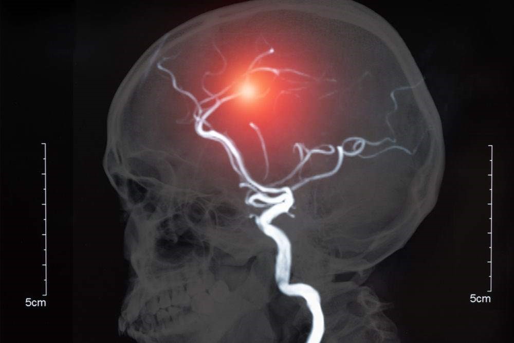
Diagnosis for unruptured cerebral aneurysms
- First level: Often CT brain scan can lead to a suspicion, but much more frequently these are occasional findings in MRI performed for other reasons.
- Second level: Angio MRI (this is a normal MRI that does not require contrast medium) and ANGIOTAC (this is a normal CT scan but requires contrast medium).
- Third level: Cerebral Angiography (local anaesthesia in the groin, catheterization through the femoral artery to reach the intracranial vessels and contrast is injected obtaining a complete dynamic visualisation of the cerebral flow) to be performed in doubtful cases or where it is necessary to know the cerebral flow and anatomical variations.
Unruptured cerebral aneurysms, treatments
It is up to the neurosurgeon to assess whether the lesion needs treatment or only observation.
At present, the literature does not provide reliable guidelines but only guidelines.
The right approach is a case-by-case assessment, taking into account the age, location of the lesion and the patient’s psychological state towards the new pathology.

It must also be considered that the treatment of a non-ruptured aneurysm presents in most cases, risks of morbidity and mortality significantly lower than subarachnoid haemorrhage, which represents an additional pathology with a whole series of complications not related to the vascular lesion, but to the blood “irritating” the cerebral surface.
The neurosurgical treatment of unruptured aneurysms is elective and presents limited risks in relation to the size of the sac, the location of the lesion and the age of the patient.
If the multidisciplinary team has given an indication for treatment, there are two possibilities:
- Microsurgical treatment
- Endovascular treatment
Endovascular treatment is not an alternative to microsurgical treatment, but a real choice of intervention.
Some aneurysms have an indication for surgery, others for endovascular treatment.
It is up to the team to assess the treatment of choice in each case.
Microsurgical treatment consists of excluding the aneurysm sac by placing one or more “clips” (small pins) at the level of the collar of the malformation.
It is performed with the aid of the most modern technology:
- Operating microscope
- Intraoperative fluoroangiography
- Intraoperative neurophysiological monitoring
- 3D endoscopy
- Intraoperative microdoppler
The risks are limited, given that the cerebral vessels rest on the surface of the brain and not inside, and that the microsurgical procedure therefore “works” on the surface without passing through the brain tissue.
The use of “intraoperative monitors” for motor and sensory assessment of the patient during the course of treatment is essential.
Endovascular treatment is a normal angiography procedure that consists of reaching the cerebral vessels through the femoral artery and filling the aneurysm sac with small titanium filaments or placing stents (small cylinders of malleable materials) that exclude the aneurysm from the brain.
The risks are related to the possibility of transient or permanent ischaemic events (higher in stents than in coils) and in the possible rupture of the aneurysm intraprocedurally.
The results of endovascular treatment may not be definitive and need serious follow-up over the years.
Prevention
There is no real prevention programme.
If a non-ruptured aneurysm is found and judged by the team to be “watchable”, it becomes imperative:
- Check blood pressure
- Stop smoking
Read Also:
Cerebral Aneurysm: What It Is And How To Treat It
Ruptured Aneurysms: What They Are, How To Treat Them
Pre-Hospital Ultrasound Assessment In Emergencies