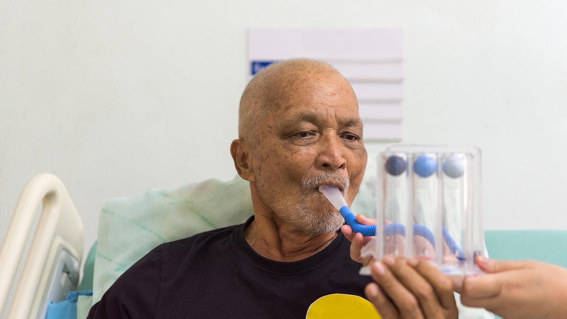
Ventilatory failure (hypercapnia): causes, symptoms, diagnosis, treatment
Hypercapnia, what causes ventilatory insufficiency? In the body, the production of the energy necessary for survival requires a constant supply of oxygen and nutrients to the tissues
Respiration provides a constant supply of oxygen to the lungs, where this gas diffuses through the alveolar-capillary membrane into the blood (external respiration).
The circulatory system then distributes the oxygenated blood to various vascular beds, where oxygen is supplied to the various tissues (internal respiration).
In addition to providing oxygenation of the blood, the lungs also serve to rid the body of carbon dioxide (CO2), a residual product of metabolism.
Carbon dioxide, carried by venous blood, diffuses into the alveoli and is subsequently exhaled into the atmosphere.
Various diseases of medical interest can lead to inadequate gas exchange and thus respiratory insufficiency, which can be ventilatory (hypercapnia) or oxygenation (hypoxaemia).
The amounts of oxygen consumed and carbon dioxide produced each minute are determined by the extent of the patient’s metabolism.
Exercise and fever are examples of factors that increase the body’s metabolism and place greater demands on the respiratory system.
When the cardio-pulmonary reserve is limited by the presence of a pathological process, fever can represent an additional stress that can precipitate respiratory failure and thus tissue hypoxia.
Ventilatory failure (hypercapnia)
In ventilatory insufficiency there is inadequate ventilation between the lungs and the atmosphere which ultimately results in an inappropriate elevation of the partial pressure of carbon dioxide in arterial blood (PaCO2) to values above 45 mmHg (hypercapnia).
Ventilatory failure (hypercapnia) is generally considered to be
- mild with PCO2 between 45 and 60 mmHg;
- moderate with PCO2 between 60 and 90 mmHg;
- severe with PCO2 above 90 mmHg.
When PCO2 exceeds 100 mmHg, coma may occur and, above 120 mmHg, death.
PCO2 is measured by haemogasanalysis.
We remind the reader that the ability to inhale requires the full efficiency of the nervous system, which must stimulate the respiratory muscles.
The contraction of the diaphragm reduces intra-thoracic pressure and causes gas to penetrate the lungs.
Minimal effort is required for this activity if the rib cage is intact, the airways pervious and the lungs distensible.
The ability to exhale, on the other hand, requires patency of the airways and lung parenchyma, which has sufficient elasticity to keep the bronchioles open until exhalation is complete.
Hypercapnia, causes and risk factors
Causes of ventilatory insufficiency include: depression of respiratory centres by pharmacological substances, brain diseases, spinal cord abnormalities, muscle diseases, rib cage abnormalities and upper and lower airway obstructions.
Upper airway obstruction can occur during acute infections and during sleep, when muscle tone is reduced.
Numerous factors can contribute to inspiratory muscle weakness and tip the balance in favour of acute ventilatory failure.
Malnutrition and electrolyte disorders can weaken the ventilatory muscles, while pulmonary hyperinflation (e.g. from pulmonary emphysema) can make the diaphragm less efficient.
Lung hyperinflation forces the diaphragm to assume an abnormally low position, which in turn leads to a mechanical disadvantage.
These problems are common in patients with acute and chronic obstructive pulmonary disease (bronchial asthma, chronic bronchitis and pulmonary emphysema).
Pathophysiology
An acute increase in PaC02 leads to a decrease in arterial blood pH.
The combination of elevated PaC02 and acidosis can have marked effects on the organism, especially when ventilatory failure is severe.
Severe acute respiratory acidosis results in impaired cognitive function due to depression of the central nervous system.
Cerebral and peripheral vessels dilate in response to hypercapnia.
Symptoms and signs
There are few clinical signs suggestive of elevated PaCO2.
Clinical signs suggestive of ventilatory failure include:
- headache;
- decreased vigilance;
- warm flushed skin;
- hypersyphilic peripheral pulses.
These findings are, however, extremely non-specific as they appear in numerous conditions other than ventilatory failure.
Since hypoxaemia is often present in a patient with ventilatory failure, it is common to observe the simultaneous appearance of signs of inadequate peripheral oxygenation.
Hypothermia and loss of consciousness are common findings, on the other hand, when ventilatory failure is the result of an overdose of substances with a sedative pharmacological effect. Sedatives and tricyclic antidepressants often result in pupillary dilation and fixation.
Tricyclic antidepressants also increase heart rate and blood pressure.
In the case of drug overdose, respiratory sounds are often evident despite the fact that aspiration has occurred.
This is more likely with sedative and alcohol abuse (as a result of the diminished swallowing reflex) and may result in rales in the right lower lobe.
Clinical signs of diaphragmatic fatigue are an early warning finding of respiratory failure in a patient with respiratory distress.
Such signs are, in fact, strongly suggestive of the need for immediate ventilatory assistance of the patient.
Diaphragm fatigue initially causes the appearance of tachypnoea, followed by periods of respiratory alternation or paradoxical abdominal breathing.
Respiratory alternation consists of the appearance of alternating for short periods of time between breathing with the accessory muscles and with the diaphragm.
Paradoxical abdominal breathing, on the other hand, is recognised on the basis of inward movement of the abdomen with each respiratory effort.
This phenomenon is due to the flaccidity of the diaphragm causing it to pull upwards whenever the accessory muscles of respiration create negative intrathoracic pressure.
Diagnosis of ventilatory failure (hypercapnia)
Anamnesis and objective examination are obviously the first steps in diagnosis.
Measurement of blood gas values is very important in assessing patients with ventilatory failure.
The severity of ventilatory failure is indicated by the extent of the increase in paCOz.
The assessment of blood pH identifies the degree of respiratory acidosis present and suggests the urgency of treatment.
The patient requires immediate treatment if the pH falls below 7.2.
Treatment
Acute elevation of arterial PCO2 indicates that the patient is unable to maintain adequate alveolar ventilation and may require ventilatory assistance.
PaCO2 does not have to exceed normal values for there to be an indication for ventilatory assistance.
For example, if the PaCO2 is 30 mmHg and then, due to respiratory muscle fatigue rises to 40 mmHg, the patient may benefit considerably from immediate intubation and mechanical ventilation.
This example therefore clearly illustrates how documenting the trend (“trending”) of arterial PaCO2 values can help in giving an indication for assisted ventilation.
Once the patient has been intubated, the set tidal volume should be 10-15 cc/Kg of ideal body weight (e.g. in obese patients a huge tidal volume is not necessary).
Current volumes below this tend to result in collapse of the more peripheral lung units (atelectasis), while current volumes above 10-15 cc/kg tend to overdistend the lungs and can cause barotrauma (pneumothorax or pneumomediastinum).
The ventilatory rate needed by the patient depends on his metabolism, although
- adult subjects usually require 8-15 respiratory acts/minute. However, ventilation is modified in most patients to maintain PaCO2 values between 35 and 45 mmHg. An exception is the patient with cerebral oedema, in whom lower PaCO2 values may prove useful in reducing intracranial pressure
- Another exception is patients with chronically high PaCO values in whom the aim of mechanical ventilation is to bring the pH back within normal limits and the patient’s PCO2 back to its baseline values. If the patient with chronic hypoventilation and CO2 retention is ventilated vigorously enough until a normal PCO2 is achieved, the problem of respiratory alkalosis arises in the short term and weaning the patient off mechanical ventilation in the long term.
The doctor should however determine the cause of the ventilatory failure before starting symptomatic treatment.
In the case of drug overdose, efforts should be made to identify the compound responsible, the amount of drug ingested, the length of time since ingestion and the presence or absence of traumatic injury.
General objectives in the treatment of logical drug overdose are to prevent absorption of the toxin (gastric lavage or stimulation of the vomiting reflex and use of activated charcoal), to increase excretion of the drug (dialysis) and to prevent accumulation of the toxic metabolic products (e.g. acetylcysteine is the antidote of choice for acetaminophen overdose).
Weaning the patient off mechanical ventilation can begin as soon as the cause of the respiratory failure has been corrected and the medically relevant clinical condition stabilised.
Weaning parameters help to define when weaning has a consistent probability of success.
Physicians should use several parameters to decide when to start weaning from ventilation, as any one of them alone can be confusing. In adult patients, the combination of a spontaneous tidal volume of more than 325 cc and a spontaneous respiratory rate of less than 38 acts/minute seems to be a good indicator of success in weaning.
Methods used in weaning include IMV, pressure support and the ‘T’ tube.
Each of these methods has advantages and disadvantages, but each should be able to effectively wean most patients as soon as possible.
Each of the methods is based on the gradual reduction of ventilatory support under controlled conditions during close monitoring of the patient.
Finally, extubation can be performed when the swallowing reflex is intact and the endotracheal tube is no longer needed.
Weaning to IMV is carried out by reducing the number of respiratory acts per minute to an interval of a few hours, until the patient no longer requires mechanical support or demonstrates poor tolerance to weaning (e.g. 20% changes in heart rate and blood pressure).
The main disadvantage of IMV is the potential increase in respiratory work imposed on the patient during spontaneous breathing (13).
This increase in work is mainly due to the excessive resistance placed on the demand valve. More recently developed ventilators, however, attempt to correct this problem.
Pressure support helps to overcome the work imposed by the resistance of the artificial circuit by administering a predetermined positive pressure during inspiration.
Weaning with pressure support requires reducing the pressure support gradually with constant monitoring of the patient’s clinical condition.
Once the patient is able to tolerate low levels of pressure support (e.g. less than 5 cm H2O) ventilatory assistance can be discontinued.
T-tube weaning is, on the other hand, performed by suspending mechanical ventilation for a short period of time and placing the patient under a continuous flow of air at a pre-determined FiO2.
The time during which the patient is allowed to breathe spontaneously is gradually extended until signs of stress appear or the subject requires mechanical ventilatory support again.
Read Also:
Emergency Live Even More…Live: Download The New Free App Of Your Newspaper For IOS And Android
Obstructive Sleep Apnoea: What It Is And How To Treat It
Obstructive Sleep Apnoea: Symptoms And Treatment For Obstructive Sleep Apnoea
Our respiratory system: a virtual tour inside our body
Tracheostomy during intubation in COVID-19 patients: a survey on current clinical practice
FDA approves Recarbio to treat hospital-acquired and ventilator-associated bacterial pneumonia
Clinical Review: Acute Respiratory Distress Syndrome
Stress And Distress During Pregnancy: How To Protect Both Mother And Child
Respiratory Distress: What Are The Signs Of Respiratory Distress In Newborns?
Respiratory Distress Syndrome (ARDS): Therapy, Mechanical Ventilation, Monitoring
Tracheal Intubation: When, How And Why To Create An Artificial Airway For The Patient
What Is Transient Tachypnoea Of The Newborn, Or Neonatal Wet Lung Syndrome?
Traumatic Pneumothorax: Symptoms, Diagnosis And Treatment
Diagnosis Of Tension Pneumothorax In The Field: Suction Or Blowing?
Pneumothorax And Pneumomediastinum: Rescuing The Patient With Pulmonary Barotrauma
ABC, ABCD And ABCDE Rule In Emergency Medicine: What The Rescuer Must Do
Multiple Rib Fracture, Flail Chest (Rib Volet) And Pneumothorax: An Overview
Internal Haemorrhage: Definition, Causes, Symptoms, Diagnosis, Severity, Treatment
Cervical Collar In Trauma Patients In Emergency Medicine: When To Use It, Why It Is Important
KED Extrication Device For Trauma Extraction: What It Is And How To Use It
How Is Triage Carried Out In The Emergency Department? The START And CESIRA Methods
Chest Trauma: Clinical Aspects, Therapy, Airway And Ventilatory Assistance


