
Vertebral fracture: causes, classification, risks, treatment, paralysis
Vertebral fracture: in medicine, the term ‘fracture’ is used to refer to the continuous breaking of a bone, usually as a result of a traumatic event whose force exceeds the bone’s resistance and it therefore ‘breaks’
Typical examples of a frequent fracture are the femur or humerus.
When a fracture affects one or more vertebrae, i.e. the bones that make up our spinal column, it is called a ‘vertebral fracture’.
All vertebrae can be affected by a fracture, so – depending on the affected area – we will have a fracture in the cervical, thoracic, lumbar, sacral and coccygeal vertebrae.
A vertebral fracture is an extremely variable event in terms of severity
Depending on the cause, the vertebrae may fracture more or less severely and – above all – may undermine the integrity of the spinal nerves and the spinal cord: in the latter case, the vertebral fracture becomes an extremely frightening event, as it may lead to permanent motor and/or sensory neurological deficits (e.g. paralysis) and, in the most serious cases, even the death of the patient.
A vertebral fracture can be related to myelopathy (bone marrow disease), radiculopathy (spinal nerve root disease) and/or discopathy (intervertebral disc disease).
Vertebral fracture classification
Vertebral fractures are the subject of numerous classifications, although currently the Denis and AO classifications are mainly used.
Denis classification
Denis divided the vertebra into three columns: anterior (vertebral body), middle (pedicles) and posterior (laminae, articular processes and spinous) with their ligaments.
According to Denis’s classification there are minor fractures, which affect the transverse and spinous processes, the laminae and the joint isthmus, and major ones:
- luxation fractures: these are accompanied by a slippage of one vertebra with respect to the other, with frequent involvement of the nerve structures housed within the spinal canal and, consequently, neurological deficits. These fractures are unstable and must always be treated surgically, with the aim of decompressing the nerve structures and stabilising the fractured section of the column by using metal synthesis means, usually in titanium alloy (pedicle screws, vertebral body substitutes);
- Compression fractures: these are fractures that occur on the basis of compressive forces that tend to cause small cracks within the vertebral bodies, resulting in sinking and loss of height of the vertebral bodies. If the loss of height caused by the deformation exceeds 50%, it is best to proceed to surgical treatment with stabilisation systems similar to those described for dislocation fractures, or with minimally invasive devices that allow the vertebral body to be reshaped and strengthened through the use of acrylic resins or synthetic bone substitutes (hydroxyapatite). If the loss of height is less than 50%, they can be treated conservatively with orthopaedic braces or consolidation techniques using percutaneous vertebroplasty. If they cause compression of the nerve structures, which is very rare, surgical decompression of the spinal canal is added to the above techniques;
- burst fractures: these consist of a multiple fragment fracture of the entire vertebral body with an axial loading mechanism leading to divergence of the pedicles and retropulsion of a bone fragment into the spinal canal. They are potentially unstable and should be treated surgically. If decompression is required, a laminectomy is performed to free the nerve structures or, if necessary, the entire vertebral body is replaced with metal prostheses inserted through anterior approaches through the chest or abdomen. If replacement of the vertebral body is not necessary, generally when the narrowing of the canal due to retropulsion of the body fragment is less than 50% of the normal antero-posterior diameter, posterior approaches using pedicle screws can be used in the traditional open technique, or percutaneous minimally invasive techniques if the situation does not require surgical decompression of the nerve structures;
- flexion/distraction fracture (or Chance fracture): these are characterised by an injury that most often extends to the anterior, middle and posterior compartments of a vertebra; in fact, therefore, in flexion/distraction vertebral fractures there is total involvement of the vertebra. Vertebral flexion/distraction fractures occur in frontal car accidents in which the person involved was wearing a lap belt. This leads to an abnormal forward displacement of the upper body, while the pelvis remains stationary on the car seat, because it is locked in place by the lap belt. A vertebral bending distraction fracture hardly ever affects stretches of the spine other than the thoracic or lumbar spine;
- transverse process fracture: these are characterised by the injury of one or more of the transverse processes present in a vertebra. A vertebral fracture of the transverse process is a stable fracture and therefore not particularly serious. Normally, episodes of vertebral fracture of the transverse process are the result of abnormal rotation or abnormal lateral bending of the vertebral column.
OA classification of vertebral fracture
The OA classification divides thoracolumbar fractures into type A (compression), type B (flexion-distraction) and type C (type B + rotational component).
This classification has further categories based on various parameters, but basically the same considerations as those discussed in Denis’ system apply.
Causes of vertebral fractures
Vertebral fractures can be of two main types:
- traumatic vertebral fractures: these are caused by trauma, which is so severe that it breaks a healthy vertebra (about 95% of all vertebral fracture cases);
- pathological vertebral fractures: the fracture occurs without any trauma or in the presence of mild trauma that would not be able to break a healthy vertebra; in this case, the vertebra is ‘sick’ and has a loss of strength (about 5% of total vertebral fracture cases).
The main causes of vertebral fracture due to trauma are:
- traffic accidents (almost half of the total cases);
- falls from heights;
- sports injuries, especially those involving physical contact, such as rugby, American football and football, but also those involving horseback riding;
- acts of violence (beatings, gunshots, etc.).
The diseases that can affect a vertebra and lead to a pathological fracture are generally of a metabolic type:
- metabolic: such as osteopenia or osteoporosis;
- neoplastic: such as tumours or bone metastases.
Risk factors
The following are at greater risk of vertebral fracture
- men (the male/female ratio is 4:1);
- young people between 18 and 25 years of age
- the elderly > 70 years old;
- women after the menopause (a period when the risk of osteoporosis increases);
- professional athletes in contact sports;
- those who ride horses or motorbikes;
- those suffering from osteopenia or osteoporosis;
- those who carry out work where there is a risk of falling (e.g. construction workers);
- those who have a primary vertebral tumour;
- those with terminal cancer with bone metastases to the vertebrae.
Vertebral fractures due to osteoporosis often recur, especially if the patient does not manage the bone-weakening condition.
Symptoms and signs of vertebral fracture
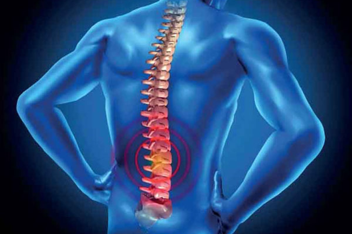
A vertebral fracture is responsible for back pain.
Sometimes moderate, sometimes intense (depending on the extent of the fracture), this pain has the particularity of worsening with movement.
If the vertebral fracture is accompanied by an injury to the spinal cord and/or spinal nerves, the symptom picture will be enriched by neurological disorders, such as
- loss of control of the anal and/or bladder sphincter;
- sense of numbness along the limbs;
- tingling along the limbs;
- sense of muscle weakness along the limbs.
It should also be noted that, in the case of vertebral fractures close to the head, the energy of the injury may spread to the brain and cause loss of consciousness.
Paralysis
One of the major risks of a vertebral fracture is damage to spinal nerves and bone marrow, which can lead to partial or total, temporary or permanent paralysis of different parts of the body depending on the site of the injury.
Below is a schematic of the possible extent of paralysis (in blue) depending on the specific site of injury.
Generally speaking, we can say that the “higher” the spinal cord damage, the more extensive the possible paralysis.
Diagnosis
Generally speaking, the following are essential for formulating the diagnosis of a vertebral fracture

- anamnesis: this consists of collecting, by means of specific questions, all the data of medical interest useful for identifying the cause and predisposing factors of a certain condition. In the case of vertebral fractures following serious trauma to the spinal column, the anamnesis is difficult to carry out because the patient is not in a position to answer. In such cases, important help may come from the person who witnessed the accident. When, on the other hand, the vertebral fracture is the result of non-weakening of the bones, the evaluation of the clinical history constitutes a fundamental step in the diagnostic pathway;
- physical examination: this consists of a careful inspection of the painful area, combined with an examination of the head, chest, abdomen, pelvis and limbs. An objective examination is unlikely to determine the type of vertebral fracture present;
- diagnostic imaging: X-ray, CT scan and magnetic resonance imaging of the spine;
- neurological examination: this allows the doctor to establish whether the vertebral fracture has in any way involved the spinal cord or neighbouring spinal nerves.
It should be noted that, when the vertebral fracture is the result of trauma that may have damaged the spinal cord, the doctor has a duty to establish the patient’s vital signs before any further assessment of the extent of the injury; carried out in an emergency, this approach protects the patient from any manoeuvres that could make the situation worse.
Therapies
The treatment of a vertebral fracture depends essentially on the type of fracture.
In general, the treatment approach is conservative in cases of mild and stable vertebral fractures, and surgical in cases of severe and unstable vertebral fractures.
At the end of the therapy aimed at repairing a vertebral fracture, a cycle of rehabilitation treatment (physiotherapy) always follows.
Conservative therapy
Conservative treatment of vertebral fractures involves the use of an orthopaedic back brace (orthosis) to keep the spine immobile and aligned during the bone healing process, and then, once the bone repair process is complete, includes a course of rehabilitation treatment (physiotherapy).
Conservative treatment is usually used in the case of:
- vertebral compression fractures;
- vertebral fractures of the transverse process.
It should be noted, however, that conservative therapy may also be used in the case of slight vertebral flexion/distraction fractures (fractures in which the injury is confined to the vertebral body).
Surgical therapy
The surgical treatment of vertebral fractures includes delicate vertebral fusion operations (in which the doctor uses screws, hooks, etc.), vertebroplasty or kyphoplasty, the aim of which is to restore the anatomy of the damaged vertebra or vertebrae and relieve any pressure on the spinal cord or neighbouring spinal nerves; as with conservative therapy, the surgical treatment of vertebral fractures also includes a course of rehabilitation.
As a rule, surgical treatment is used in the case of:
- Vertebral burst fractures;
- vertebral fractures due to bending/distraction with total vertebral involvement;
- vertebral fractures with dislocation.
Readers are reminded that, unfortunately, spinal cord injuries are permanent, which means that there is no medical or surgical therapy to repair them.
Physiotherapy
After a spinal fracture, physical rehabilitation (or physiotherapy) is used to restore the elasticity and muscle tone of the back that existed before the injury.
The duration of physiotherapy varies depending on the type of vertebral fracture.
Complications of vertebral fracture therapy
The treatment of vertebral fractures forces the patient to rest in bed; the resulting immobility is a dangerous factor in promoting the phenomenon of venous thrombosis along the limbs, especially the lower ones.
In addition to this dangerous complication, there are also possible complications that may arise from the use of surgery and which characterise this therapeutic option, regardless of the field of application (such complications consist of infections, haemorrhages, etc.).
Prognosis
Generally speaking, the more severe a vertebral fracture is, the less chance there is of a complete recovery; even, in the case of vertebral fractures associated with spinal cord injury, the restoration of certain motor functions (including control of the kidney and bladder sphincters) is impossible.
Bone repair times
In the case of a vertebral fracture, bone repair times are between 6 and 12 weeks.
Read Also:
Emergency Live Even More…Live: Download The New Free App Of Your Newspaper For IOS And Android
Bone Cysts In Children, The First Sign May Be A ‘Pathological’ Fracture
Fracture Of The Wrist: How To Recognise And Treat It
Fractures Of The Growth Plate Or Epiphyseal Detachments: What They Are And How To Treat Them
Stress Fractures: Risk Factors And Symptoms
Calcaneal Fractures: What They Are, How To Intervene
Greenstick Fractures: What They Are, What The Symptoms Are And How To Treat Them
Broken Bone First Aid: How To Recognise A Fracture And What To Do
Fractures Of The Upper Limb: What It Looks Like And How To Deal With A Broken Arm
Understanding Bone Fractures: Diagnosis And Treatment
Facial Trauma With Skull Fractures: Difference Between LeFort Fracture I, II And III