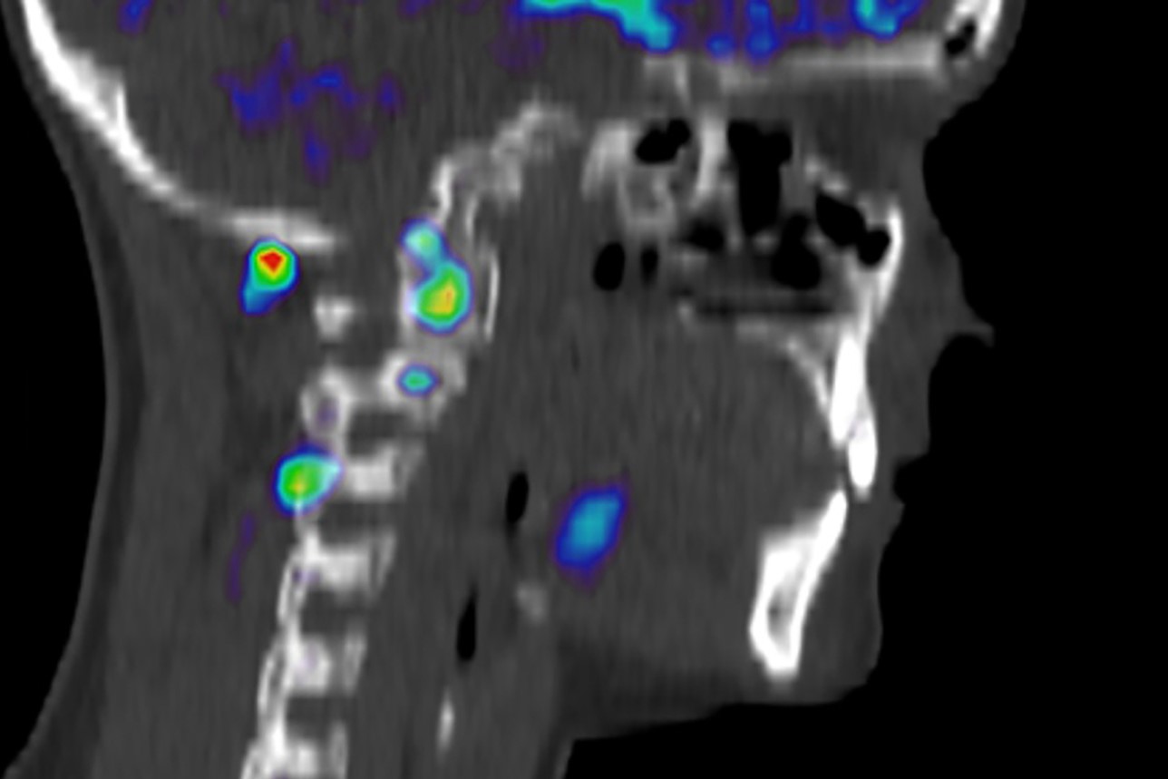
Visualizing Pain: injuries from whiplash made visible with new scanning approach
Whiplash, a condition often associated with rear-end motor vehicle collisions, is notoriously difficult to diagnose and treat
There are hundreds of thousands of whiplash injuries due to car accidents reported worldwide each year, causing significant societal and financial costs
Until now, specific diagnosis of areas of the body affected by whiplash has been elusive because injuries associated with the syndrome do not appear on standard scans.
Now, an international team led by researchers from the Harvard Medical School Department of Physical Medicine and Rehabilitation at Spaulding Rehabilitation Hospital has used a new approach to scanning that allowed them to capture images of injuries from whiplash.
The researchers said that this new approach, which shows areas of inflammation from whiplash injuries, will allow clinicians to better target medical treatments for whiplash
Their findings were published online July 2 in the journal PAIN.
Imaging musculoskeletal images is important because it allows clinicians to know precisely where an injury is located and can give important information about the intensity and extent of the injury, the researchers said.
“An objective visualization and quantification of the injury and possible inflammation in whiplash-associated disorders would support a better diagnosis, strengthen patients’ subjective report of pain, and assist clinical decisions,” said study lead author Clas Linnman, HMS assistant professor of physical medicine and rehabilitation at the Spaulding Neuroimaging Lab.
THE BEST SPINE BOARDS? VISIT THE SPENCER BOOTH AT EMERGENCY EXPO
Linnman noted that the difficulties of detecting and diagnosing lesions related to the experienced pain reported by people with whiplash, together with the lack of an accepted concept for what causes the symptoms in whiplash-associated disorders, contribute to considerable personal, societal, and economic problems
For this study, 16 young adult patients with whiplash injury grade II were recruited at the emergency department and underwent PET/CT scans at the time of their initial hospital visit and at a follow-up six months after injury.
For the PET/CT scans, the researchers used a special tracer, [11C]D-deprenyl, that has been shown to help visualize inflammation in other types of musculoskeletal injuries.
Eight healthy individuals were also imaged as a control group.
Subjective pain levels, self-rated neck disability, and active cervical range of motion were recorded at the two imaging sessions.
The results showed that the molecular aspects of inflammation and possible tissue injuries in muscles and facet joints after acute whiplash injury could be visualized, objectively quantified, and followed over time with [11C]D-deprenyl PET/CT.
The new approach allowed the researchers to see which peripheral structures were affected in whiplash injury, which strengthens the idea that PET detectable organic lesions in peripheral tissue are relevant for the development of persistent pain and disability in whiplash injury, the researchers said.
“It is our hope that this work will be an important contribution to better understanding chronic musculoskeletal pain conditions,” Linnman said.
This study was supported by the LF Insurance Research Foundation, the Berzelii Technology Centre for Neurodiagnostics (grant 29797-1), the Swedish Medical Research Council (Grant 9459), and the Scott Schoen and Nancy Adams Discovery Center for Recovery from Chronic Pain at Spaulding Rehab Hospital. All authors declare that they have no conflict of interest.
Whiplash_injuries_associated_with_experienced_pain.97970Read Also:
Burnout In Paramedics: Exposure To Critical Injuries Among Ambulance Workers In Minnesota
Spinal Column Injuries, The Value Of The Rock Pin / Rock Pin Max Spine Board


