
What is Acute Lymphocytic Leukemia?
Acute lymphocytic leukemia (ALL), also known as acute lymphoblastic leukemia, refers to an abnormal growth of lymphocyte precursors or lymphoblasts
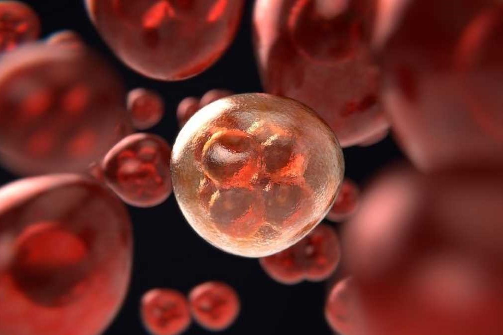
Acute leukemias have large numbers of immature leukocytes and overproduction of cells in the blast stage of maturation.
Acute lymphocytic leukemia (ALL), also known as acute lymphoblastic leukemia, refers to an abnormal growth of lymphocyte precursors or lymphoblasts
Acute leukemia is a malignant proliferation of white blood cell precursors in bone marrow or lymph tissue, and their accumulation in peripheral blood, bone marrow, and body tissues.
About 20% of leukemias are acute.
Pathophysiology
Pathogenesis isn’t clearly understood, but the pathophysiology may be explained by the following:
- Accumulation. Due to the precipitating factors, immature, non-functioning WBCs appear to accumulate first in the tissue where they originate (lymphocytes in lymph tissue, granulocytes in bone marrow).
- Infiltration. These immature WBCs then spill into the bloodstream and from there infiltrate other tissues.
- Malfunction. Eventually, this infiltration results in organ malfunction because of encroachment and hemorrhage.
One of the most common forms of acute leukemia is acute lymphocytic leukemia
Acute lymphocytic leukemia is more common in males than in females, in whites (especially in people of Jewish descent), in children (between ages 2 and 5), and in people who live in urban and industrialized areas.
- 80% of all leukemias between 2 and 5 years old are ALL.
- Acute leukemias account for 20% of adult leukemias.
Among children, however, it is the most common form of cancer.
Incidence is 6 out of every 100, 000 people.
Causes
Research on predisposing factors isn’t conclusive but points to some combination of viruses, immunologic factors, genetic factors, and exposure to radiation and certain chemicals.
- Congenital disorders. Down syndrome, Bloom syndrome, Fanconi anemia, congenital agammaglobulinemia, and ataxia-telangiectasia usually predisposes to ALL.
- Familial tendency. Genetics also play a part in the development of ALL.
- Viruses. Viral remnants have been found in leukemic cells, so they are likely one of the causes of ALL.
Signs of acute lymphocytic leukemia may be gradual or abrupt
- High fever. High fever accompanied by thrombocytopenia and abnormal bleeding (such as nosebleeds and gingival bleeding) manifests in the patient.
- Bruising. Easy bruising after minor trauma is a sign of leukemia.
- Dyspnea. A decrease in the mature blood components leads to dyspnea.
- Anemia. Anemia is present in ALL because of a decrease in mature RBCs.
- Fatigue. The patient experiences fatigue more frequently than normal.
- Tachycardia. As the oxygen-carrying component of the blood decreases, the body compensates by pumping out blood faster than normal.
Complications
Untreated, acute leukemia is invariably fatal, usually because of complications that result from leukemic cell infiltration of the bone marrow and vital organs.
- Infection. Immature WBCs are not fit to defend the body against pathogens, so infection is always a possible complication to watch out for.
- Organ malfunction. Encroachment or hemorrhage occurs when immature WBCs spill into the bloodstream and other tissues and eventually lead to organ or tissue malfunction.
The diagnosis of ALL can be confirmed with a combination of the following:
- Bone marrow aspiration. Typical clinical findings and bone marrow aspirate showing a proliferation of immature WBCs confirm ALL.
- Bone marrow biopsy. A bone marrow biopsy, usually of the posterior superior iliac spine, is part of the diagnostic workup.
- Blood counts. Blood counts show severe anemia, thrombocytopenia, and neutropenia.
- Differential leukocyte count. Differential leukocyte count determines cell type.
- Lumbar puncture. Lumbar puncture detects meningeal involvement.
- Uric acid levels. Elevated uric acid levels and lactic dehydrogenase levels are commonly found.
Medical Management
With treatment, the prognosis varies.
- Systemic chemotherapy. Systemic chemotherapy aims to eradicate leukemic cells and induce remission (less than 5% of blast cells in the marrow and peripheral blood are normal).
- Radiation therapy. Radiation therapy is given for testicular infiltrations.
- Platelet transfusion is performed to prevent bleeding and RBC transfusion to prevent anemia.
ALL (Acute lymphocytic leukemia)chemotherapy includes the following drugs and also other drugs included in the treatment:
- Vincristine. Vincristine is an anti-cancer (antineoplastic or cytotoxic) chemotherapy drug and is classified as a plant alkaloid.
- Prednisone. This drug works is by altering the body’s normal immune system responses.
- Cytarabine. Cytarabine belongs to the category of chemotherapy called antimetabolites, wherein When the cells incorporate these substances into the cellular metabolism, they are unable to divide and they attack cells at very specific phases in the cycle.
- L-asparaginase. Asparaginase breaks down asparagine in the body, so since the cancer cells cannot make more asparagine, they die.
- Daunorubicin. Daunorubicin is classified as an antitumor antibiotic which is made from natural products produced by species of the soil fungus Streptomyces, and these drugs act during multiple phases of the cell cycle and are considered cell-cycle specific.
- Antibiotic, antifungal, and antivirals. These control infection, a common complication of acute leukemias.
Surgical Management
Aggressive treatment may include surgical management through:
- Bone marrow transplant. Bone marrow transplant is a choice that can be considered for a patient with ALL.
- Stem cell transplant. Stem cell transplant in ALL is one of the latest development in the treatment of acute leukemias
Nursing Management
The care plan for the leukemic patient should emphasize comfort, minimize the adverse effects of chemotherapy, promote preservation of veins, manage complications, and provide teaching and psychological support.
Nursing Assessment
The clinical picture varies with the type pf leukemia as well as the treatment implemented, so the following must be assessed:
- Health history. The health history may reveal a range of subtle symptoms reported by the patient before the problem is detectable on physical examination.
- Physical examination. A thorough, systematic assessment incorporating all body systems is essential.
- Laboratory results. The nurse also must closely monitor the results of laboratory studies and culture results need to be reported immediately.
Nursing Diagnosis
Based on the assessment data, major nursing diagnoses for the patient with ALL may include:

- Risk for infection related to overproduction of immature WBCs.
- Risk for impaired skin integrity related to toxic effects of chemotherapy, alteration in nutrition, and impaired immobility.
- Imbalanced nutrition, less than body requirements, related to hypermetabolic state, anorexia, mucositis, pain, and nausea.
- Acute pain and discomfort related to mucositis, leukocyte infiltration of systemic tissues, fever, and infection.
- Hyperthermia related to tumor lysis or infection.
- Fatigue and activity intolerance related to anemia, infection, and deconditioning.
Read Also
Emergency Live Even More…Live: Download The New Free App Of Your Newspaper For IOS And Android
Leukaemia: Symptoms, Causes And Treatment
Leukaemia: The Types, Symptoms And Most Innovative Treatments
Lymphoma: 10 Alarm Bells Not To Be Underestimated
Non-Hodgkin’s Lymphoma: Symptoms, Diagnosis And Treatment Of A Heterogeneous Group Of Tumours
CAR-T: An Innovative Therapy For Lymphomas
Acute Lymphoblastic Leukaemia: Long-Term Outcomes Described For Childhood ALL Survivors
Colour Changes In The Urine: When To Consult A Doctor
Why Are There Leukocytes In My Urine?
Acute Lymphocytic Leukaemia: What Is It?
Rectal Cancer: The Treatment Pathway
Testicular Cancer And Prevention: The Importance Of Self-Examination
Testicular Cancer: What Are The Alarm Bells?
The Reasons For Prostate Cancer
Bladder Cancer: Symptoms And Risk Factors
Breast Cancer: Everything You Need To Know
Rectosigmoidoscopy And Colonoscopy: What They Are And When They Are Performed
Bone Scintigraphy: How It Is Performed
Fusion Prostate Biopsy: How The Examination Is Performed
CT (Computed Axial Tomography): What It Is Used For
What Is An ECG And When To Do An Electrocardiogram
Positron Emission Tomography (PET): What It Is, How It Works And What It Is Used For
Single Photon Emission Computed Tomography (SPECT): What It Is And When To Perform It
Instrumental Examinations: What Is The Colour Doppler Echocardiogram?
Coronarography, What Is This Examination?
CT, MRI And PET Scans: What Are They For?
MRI, Magnetic Resonance Imaging Of The Heart: What Is It And Why Is It Important?
Urethrocistoscopy: What It Is And How Transurethral Cystoscopy Is Performed
What Is Echocolordoppler Of The Supra-Aortic Trunks (Carotids)?
Surgery: Neuronavigation And Monitoring Of Brain Function
Robotic Surgery: Benefits And Risks
Refractive Surgery: What Is It For, How Is It Performed And What To Do?
Anorectal Manometry: What It Is Used For And How The Test Is Performed