
What is digital mammography and what advantages it has
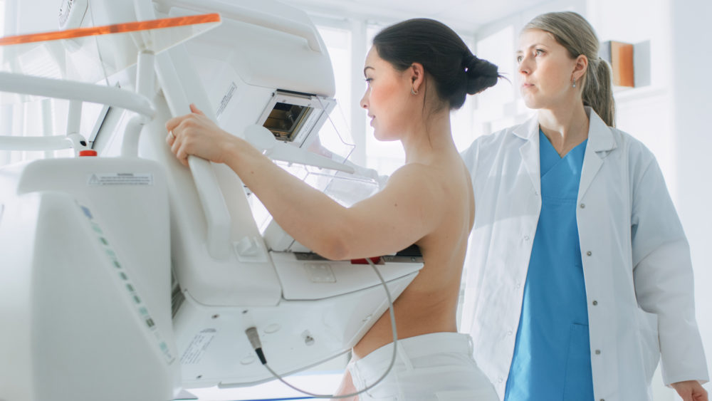
Digital mammography is a new diagnostic method that uses a piece of equipment called a digital mammograph to form the mammography image
In digital mammography, the X-ray film is replaced by a detector
This absorbs the X-rays transmitted through the breast and converts their energy into electronic signals, which are digitised and fixed in the computer’s memory.
An image, the digital mammogram, is then taken from this data and displayed on a high-definition monitor.
From there, after being appropriately processed, it can be printed on film by a laser printer or stored in one of the various archiving systems available today, including CD-ROM.
The advantages of digital mammography
The traditional mammography image is a film image that, like a photograph, is no longer editable after being produced.
In addition, the breast is made up of areas of different density: since these areas are reproduced in a single image, there will be areas that are well represented, and therefore well studyable, alongside areas that are not correctly represented, too light or too dark, and therefore not correctly studyable.
The digital image, on the other hand, can also be processed by the computer after training: it can then be suitably modified by varying the parameters of contrast, brightness, magnification, etc., thus making it possible to correctly visualise each different area of the breast.
The digital image can be displayed on high-definition monitors or printed on film using laser printers.
One of the factors that sometimes prevent mammography from diagnosing a tumour is that the pathological area has too little intrinsic contrast difference to the surrounding healthy tissue.
Since the digital image can be processed after acquisition, contrast differences can be enhanced, making diagnosis easier.
The overall performance of the system, particularly in terms of contrast resolution, is significantly higher than the conventional system.
This allows images of excellent diagnostic quality to be obtained with a lower radiation dose
In addition, since the images can be reprocessed by computer, it is possible to have a good mammogram even under less than optimal exposure conditions.
This reduces the problem, common with traditional techniques, of repeating non-diagnostic tests because they are not properly exposed.
The radiation dose administered to women is thus reduced, a factor that is particularly important in mammography since the investigation, to be effective for prevention purposes, must be repeated periodically every 1-2 years.
The availability of images in digital form allows the creation of complete computerised archives, including both all clinical information about the patients and the relevant images.
A complete computerised medical record should therefore be obtained, with advantages not only for practical management but also for research and teaching aspects.
Finally, the digital image can be transmitted remotely (other workstations at hospitals, general practitioners, research centres, any computer connected by network or telephone line) with several possible applications: transmission from the place of execution to the place of reporting, transmission to reference centres for consultation, etc.
Read Also
Emergency Live Even More…Live: Download The New Free App Of Your Newspaper For IOS And Android
Breast Cancer: For Every Woman And Every Age, The Right Prevention
Transvaginal Ultrasound: How It Works And Why It Is Important
Pap Test, Or Pap Smear: What It Is And When To Do It
Mammography: A “Life-Saving” Examination: What Is It?
Breast Cancer: Oncoplasty And New Surgical Techniques
Gynaecological Cancers: What To Know To Prevent Them
Ovarian Cancer: Symptoms, Causes And Treatment

What Are The Risk Factors For Breast Cancer?
Breast Cancer Women ‘Not Offered Fertility Advice’
Ethiopia, The Minister Of Health Lia Taddesse: Six Centers Against Breast Cancer
Breast Self-Exam: How, When And Why
Fusion Prostate Biopsy: How The Examination Is Performed
CT (Computed Axial Tomography): What It Is Used For
What Is An ECG And When To Do An Electrocardiogram
MRI, Magnetic Resonance Imaging Of The Heart: What Is It And Why Is It Important?
Mammary MRI: What It Is And When It Is Done
What Is Needle Aspiration (Or Needle Biopsy Or Biopsy)?
Positron Emission Tomography (PET): What It Is, How It Works And What It Is Used For
CT, MRI And PET Scans: What Are They For?
MRI, Magnetic Resonance Imaging Of The Heart: What Is It And Why Is It Important?
Urethrocistoscopy: What It Is And How Transurethral Cystoscopy Is Performed
What Is Echocolordoppler Of The Supra-Aortic Trunks (Carotids)?
Surgery: Neuronavigation And Monitoring Of Brain Function
Robotic Surgery: Benefits And Risks
Refractive Surgery: What Is It For, How Is It Performed And What To Do?
Single Photon Emission Computed Tomography (SPECT): What It Is And When To Perform It
What Is An ECG And When To Do An Electrocardiogram
MRI, Magnetic Resonance Imaging Of The Heart: What Is It And Why Is It Important?
Mammary MRI: What It Is And When It Is Done
Mammography: How To Do It And When To Do It
Pap Test: What Is It And When To Do It?