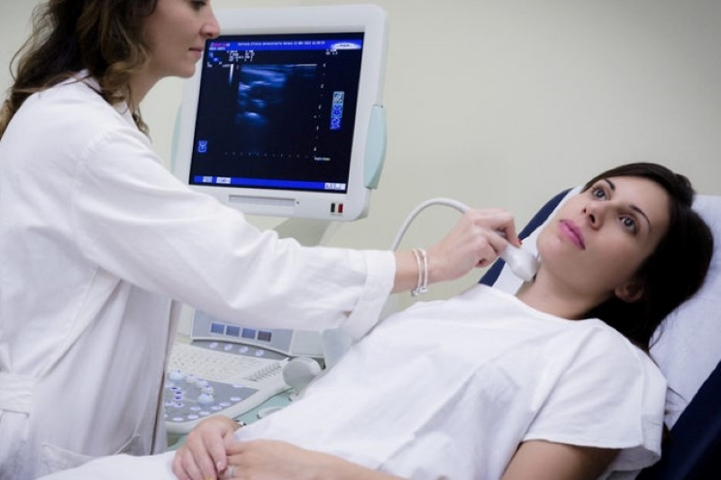
What is echocolordoppler of the supra-aortic trunks (carotids)?
Echocolordoppler of the supra-aortic trunks, also known as the carotid or epiaortic vessels, is a noninvasive diagnostic examination dedicated to the carotid and vertebral arteries, i.e., those arteries whose function is to carry blood to the brain
It is an ultrasound enriched with visual and acoustic (color-doppler) values to monitor the arterial circulation directed toward the brain, to evaluate the blood vessels and the blood flow within them, and to diagnose the possible presence of changes in the vessel walls.
What is the purpose of supra-aortic trunk echocolordoppler?
With supra-aortic trunks echocolordoppler, it is possible to establish, study or rule out the presence of plaques in the vessels, which can lead to the presence of stenosis, i.e., points where the artery, reduced in caliber, allows less blood to pass through.
This condition, by reducing the supply of oxygen to the brain, is particularly dangerous as it can promote the onset of permanent ischemia, i.e., ICTUS or a transient ischemic attack (TIA).
Thanks to the examination, it is possible to identify the site of stenosis, quantify the percentage of narrowing of the vessel, the alteration on blood flow, and, thanks to the evaluation of the vessel’s inner wall, the degree of risk of the onset of ischemic-type cerebrovascular disease even in patients who do not have stenosis.
In short, therefore, the diagnostic test is the best method for assessing the risk of cardiovascular system disease in both symptomatic and non-symptomatic subjects.
When is supra-aortic trunk echocolordoppler performed?
Supra-aortic trunks echocolordoppler, abbreviated TSA, may be requested by a physician to screen a patient following a stroke or transient cerebral ischemia, to diagnose an aneurysm, or to rule out the presence of venous thrombosis.
In addition, it can be recommended for screening patients with cardiovascular risk factors, i.e., subjects with hypertension, diabetes, hypercholesterolemia, dyslipidemia, obesity, etc.
In addition, the examination is recommended for screening of:
- patients with a family history of cerebral or cardiac ischemic events;
- smokers;
- patients about to undergo major vascular surgery;
- patients undergoing radiation therapy of the neck.
How is the supra-aortic trunk echocolordoppler performed?
The examination consists of a simple and absolutely noninvasive method.
In fact, it involves no preparation for the patient and has no contraindications.
It is performed like any ultrasound and lasts an average of 20 minutes.
At the end of the echocolordoppler examination of the sovaortic trunks, should the presence of stenosis or the risk of cerebrovascular disease be identified, it will be possible to make therapeutic decisions aimed at:
- correct risk factors through careful planning of prophylaxis suitable for slowing the progression of the disease;
- establish appropriate treatment.
In the case of the presence of stenosing or occlusive pathology affecting the supra-aortic trunks, the physician may prescribe the patient either pharmacological therapy or surgical therapy, which consists of carotid endarterectomy, aimed at removing the plaque attached to the inner surface of the vessel.
*This is indicative information; therefore, it is necessary to contact the facility where the examination is performed to obtain specific information on the preparation procedure.
Read Also
Emergency Live Even More…Live: Download The New Free App Of Your Newspaper For IOS And Android
What Is The Loop Recorder? Discovering Home Telemetry
Cardiac Holter, The Characteristics Of The 24-Hour Electrocardiogram
Peripheral Arteriopathy: Symptoms And Diagnosis
Endocavitary Electrophysiological Study: What Does This Examination Consist Of?
Cardiac Catheterisation, What Is This Examination?
Echo Doppler: What It Is And What It Is For
Transesophageal Echocardiogram: What Does It Consist Of?
Venous Thrombosis: From Symptoms To New Drugs
Heart Murmur: What Is It And What Are The Symptoms?
Cardiopulmonary Resuscitation Manoeuvres: Management Of The LUCAS Chest Compressor
Supraventricular Tachycardia: Definition, Diagnosis, Treatment, And Prognosis
Identifying Tachycardias: What It Is, What It Causes And How To Intervene On A Tachycardia
Tachycardia: Is There A Risk Of Arrhythmia? What Differences Exist Between The Two?
Do You Have Episodes Of Sudden Tachycardia? You May Suffer From Wolff-Parkinson-White Syndrome (WPW)
Transient Tachypnoea Of The Newborn: Overview Of Neonatal Wet Lung Syndrome
Paediatric Toxicological Emergencies: Medical Intervention In Cases Of Paediatric Poisoning
Valvulopathies: Examining Heart Valve Problems
What Is The Difference Between Pacemaker And Subcutaneous Defibrillator?
Heart Disease: What Is Cardiomyopathy?
Inflammations Of The Heart: Myocarditis, Infective Endocarditis And Pericarditis
Heart Murmurs: What It Is And When To Be Concerned
Broken Heart Syndrome Is On The Rise: We Know Takotsubo Cardiomyopathy
Cardiomyopathies: What They Are And What Are The Treatments
Alcoholic And Arrhythmogenic Right Ventricular Cardiomyopathy
Difference Between Spontaneous, Electrical And Pharmacological Cardioversion
What Is Takotsubo Cardiomyopathy (Broken Heart Syndrome)?
Dilated Cardiomyopathy: What It Is, What Causes It And How It Is Treated
Heart Pacemaker: How Does It Work?
Basic Airway Assessment: An Overview
Assessment Of Abdominal Trauma: Inspection, Auscultation And Palpation Of The Patient
Pain Assessment: Which Parameters And Scales To Use When Rescuing And Treating A Patient
Airway Management After A Road Accident: An Overview
Tracheal Intubation: When, How And Why To Create An Artificial Airway For The Patient
What Is Traumatic Brain Injury (TBI)?
Acute Abdomen: Meaning, History, Diagnosis And Treatment
Poison Mushroom Poisoning: What To Do? How Does Poisoning Manifest Itself?
Chest Trauma: Clinical Aspects, Therapy, Airway And Ventilatory Assistance
The Quick And Dirty Guide To Pediatric Assessment
EMS: Pediatric SVT (Supraventricular Tachycardia) Vs Sinus Tachycardia


