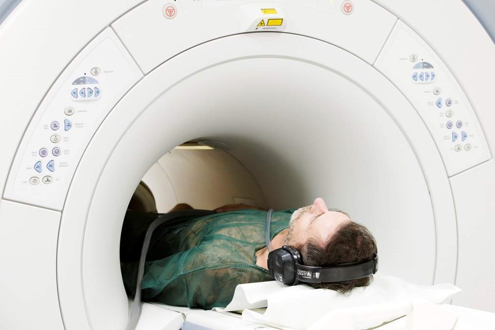
What is Magnetic resonance imaging of the heart with contrast medium (Cine MRI) and why is it performed?
Magnetic resonance imaging of the heart, also known as Cine MRI, is a diagnostic examination that allows the heart muscle in motion to be analysed in detail; thanks to a special apparatus, dynamic images can be obtained, which are then processed in the form of videos
Depending on requirements, this procedure can be performed either with or without contrast medium.
Cine MRI makes it possible to examine the anatomy of the heart and its functioning, in order to detect any abnormalities, malformations and disorders: it is one of the most reliable procedures for studying cardiac volumes, mass and function; it also makes it possible to check the atrial cavities, the functioning of the heart valves, the blood vessels responsible for myocardial blood supply and their flow rate.
Why MRI of the heart with contrast medium (Cine MRI) is performed
Magnetic resonance imaging of the heart is a specialist radiological examination that provides highly detailed information about the morphology and function of the heart muscle.
It makes it possible to analyse the health of the heart in detail, without resorting to invasive procedures; in particular, this examination is indicated for the diagnosis and monitoring of diseases and conditions, such as
- congenital malformations or acquired heart defects
- tumours affecting the heart;
- particular forms of arrhythmias
- primary or secondary cardiomyopathies;
- fibrosis of the myocardium;
- damage assessment following myocardial necrosis or infarction;
- pathologies and inflammations of the pericardium (pericarditis);
- diseases and dysfunctions of the aorta;
- valvulopathies or defects in the heart valve system;
- coronary artery dysfunctions or malformations.
How should I prepare for MRI of the heart with contrast medium (Cine MRI)
MRI of the heart requires no special preparation.
Prior to the procedure, the patient undergoes a thorough history to detect any allergies, a thorough physical examination, including a cardiological examination and an analysis of renal function.
On the day of the procedure, if contrast medium is to be used, a fast of at least 4-6 hours is recommended; if Cine MRI also includes the study of perfusion under pharmacological stress, the patient must abstain from taking excitatory substances in the preceding 12 hours and, on the doctor’s advice, it may be necessary to discontinue any pharmacological therapies beforehand.
Prior to the examination, the patient is also asked to remove any metal objects, make-up or contact lenses.
In addition, although in most cases it is not a problem, it is good practice to inform healthcare professionals if you have undergone surgery with coronary stents, if you have post-cardiac sternal sutures or if you have implants and prostheses.
Cine MRI cannot, however, be performed by patients with pacemakers or magnetically activated devices; it is also not recommended during the first trimester of pregnancy.
In general, it is a non-invasive procedure, with the exception of the intravenous administration of contrast medium, which is not painful and carries no particular risk; the only discomfort the patient may feel is caused by the noise of the instrument in operation.
*This is indicative information: it is therefore necessary to contact the facility where the examination is being performed to obtain specific information on the preparation procedure.
Read Also:
Emergency Live Even More…Live: Download The New Free App Of Your Newspaper For IOS And Android
Endocavitary Electrophysiological Study: What Does This Examination Consist Of?
Head Up Tilt Test, How The Test That Investigates The Causes Of Vagal Syncope Works
What Is Ischaemic Heart Disease And Possible Treatments
Percutaneous Transluminal Coronary Angioplasty (PTCA): What Is It?
Ischaemic Heart Disease: What Is It?
EMS: Pediatric SVT (Supraventricular Tachycardia) Vs Sinus Tachycardia
Paediatric Toxicological Emergencies: Medical Intervention In Cases Of Paediatric Poisoning
Valvulopathies: Examining Heart Valve Problems
What Is The Difference Between Pacemaker And Subcutaneous Defibrillator?
Heart Disease: What Is Cardiomyopathy?
Inflammations Of The Heart: Myocarditis, Infective Endocarditis And Pericarditis
Heart Murmurs: What It Is And When To Be Concerned
Clinical Review: Acute Respiratory Distress Syndrome
Botallo’s Ductus Arteriosus: Interventional Therapy
Heart Valve Diseases: An Overview
Cardiomyopathies: Types, Diagnosis And Treatment
First Aid And Emergency Interventions: Syncope
Tilt Test: What Does This Test Consist Of?
Cardiac Syncope: What It Is, How It Is Diagnosed And Who It Affects
New Epilepsy Warning Device Could Save Thousands Of Lives
Understanding Seizures And Epilepsy
First Aid And Epilepsy: How To Recognise A Seizure And Help A Patient
Neurology, Difference Between Epilepsy And Syncope
Positive And Negative Lasègue Sign In Semeiotics
Wasserman’s Sign (Inverse Lasègue) Positive In Semeiotics
Positive And Negative Kernig’s Sign: Semeiotics In Meningitis
Lithotomy Position: What It Is, When It Is Used And What Advantages It Brings To Patient Care
Trendelenburg (Anti-Shock) Position: What It Is And When It Is Recommended
Prone, Supine, Lateral Decubitus: Meaning, Position And Injuries
Stretchers In The UK: Which Are The Most Used?
Does The Recovery Position In First Aid Actually Work?
Reverse Trendelenburg Position: What It Is And When It Is Recommended
Drug Therapy For Typical Arrhythmias In Emergency Patients
Canadian Syncope Risk Score – In Case Of Syncope, Patients Are Really In Danger Or Not?


