What is the Loop Recorder? Discovering Home Telemetry
The loop recorder is an implantable device used for the purpose of detecting occasional heart rhythm abnormalities
The device has a cyclic memory and continuously records the heart rhythm, returning an electrocardiographic trace that can be remotely consulted and interpreted by the physician.
Unlike the Holter ECG, which allows continuous recording for 24-48 hours, loop recorders allow much longer monitoring
The device is the size of a USB stick and is placed on the chest, under the patient’s skin, through an incision.
The latest models are so small that they are called ‘injectable loop recorders’ and, once inserted, are completely invisible.
How does the Loop Recorder work?
The loop recorder is equipped with a retrospective memory, thanks to which it is possible to record the ECG trace continuously and to store only certain recording segments afterwards.
The storage can be done either by activating the recording manually using a special remote control, in cases where symptoms are present, or automatically, in cases where the device detects arrhythmias.
The length of the ECG trace can vary depending on the device, as can the internal storage capacity.
Once exhausted, the oldest traces will have to be deleted so that new ones can be recorded.
For whom is Loop Recorder recommended?
The Loop Recorder is recommended for patients who present with symptoms compatible with an arrhythmogenic disease, but which, due to their infrequency, cannot be analysed using other diagnostic tests.
In addition, the device is recommended in cases in which the patient cannot activate the recording manually or if one wishes to assess the presence of asymptomatic arrhythmias.
More specifically, the use of the device can be applied in cases of:
- recurrent loss of consciousness
- epilepsy;
- asymptomatic recurrent syncopes;
- atrial fibrillation;
- palpitations;
- cryptogenic stroke of uncertain origin.
Its use allows the physician to provide a systemic screening of possible cardiac abnormalities, to make a correct diagnosis, but also to assess the effectiveness of the patient’s anti-arrhythmic therapy.
How is implantation performed?
Implantation of the loop recorder is performed during a short Day Hospital stay, at which the patient must have been fasting for at least 8 hours.
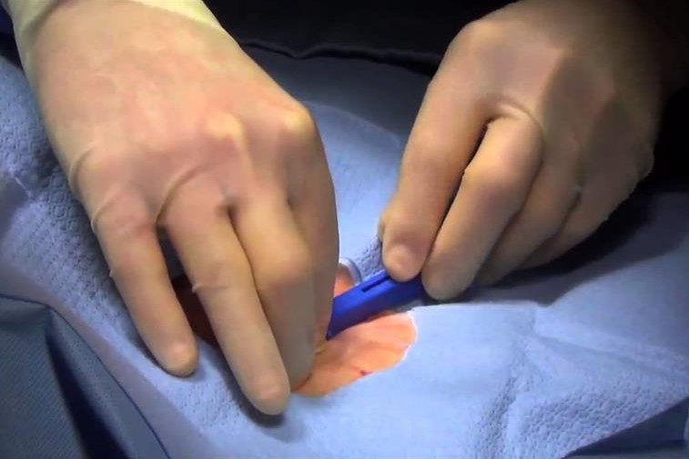
The procedure involves grafting the device under the skin through a small incision in the pectoral skin, performed under local anaesthesia.
At the end of the procedure, the incision is closed with a few resorbable stitches.
The newer, so-called injectable models, on the other hand, can be inserted via a special subcutaneous insertion system.
Once the implant has been performed, the patient must remain under observation for 1-2 hours, after which he or she can be discharged.
Complications due to implantation are usually very rare and are limited to slight soreness and a small localised haematoma in the area of implantation, which tends to recede within a few days after the procedure.
In very rare cases wound infection may occur.
*This is only indicative information: it is therefore necessary to contact the facility where the examination is being performed to obtain specific information on the preparation procedure.
Read Also:
Emergency Live Even More…Live: Download The New Free App Of Your Newspaper For IOS And Android
Cardiac Holter, The Characteristics Of The 24-Hour Electrocardiogram
Peripheral Arteriopathy: Symptoms And Diagnosis
Endocavitary Electrophysiological Study: What Does This Examination Consist Of?
Head Up Tilt Test, How The Test That Investigates The Causes Of Vagal Syncope Works
What Is Ischaemic Heart Disease And Possible Treatments
Percutaneous Transluminal Coronary Angioplasty (PTCA): What Is It?
Ischaemic Heart Disease: What Is It?
EMS: Pediatric SVT (Supraventricular Tachycardia) Vs Sinus Tachycardia
Paediatric Toxicological Emergencies: Medical Intervention In Cases Of Paediatric Poisoning
Valvulopathies: Examining Heart Valve Problems
What Is The Difference Between Pacemaker And Subcutaneous Defibrillator?
Heart Disease: What Is Cardiomyopathy?
Inflammations Of The Heart: Myocarditis, Infective Endocarditis And Pericarditis
Heart Murmurs: What It Is And When To Be Concerned
Clinical Review: Acute Respiratory Distress Syndrome
Botallo’s Ductus Arteriosus: Interventional Therapy
Heart Valve Diseases: An Overview
Cardiomyopathies: Types, Diagnosis And Treatment
First Aid And Emergency Interventions: Syncope
Tilt Test: What Does This Test Consist Of?
Cardiac Syncope: What It Is, How It Is Diagnosed And Who It Affects
New Epilepsy Warning Device Could Save Thousands Of Lives
Understanding Seizures And Epilepsy
First Aid And Epilepsy: How To Recognise A Seizure And Help A Patient
Neurology, Difference Between Epilepsy And Syncope
Positive And Negative Lasègue Sign In Semeiotics
Wasserman’s Sign (Inverse Lasègue) Positive In Semeiotics
Positive And Negative Kernig’s Sign: Semeiotics In Meningitis
Lithotomy Position: What It Is, When It Is Used And What Advantages It Brings To Patient Care
Trendelenburg (Anti-Shock) Position: What It Is And When It Is Recommended
Prone, Supine, Lateral Decubitus: Meaning, Position And Injuries
Stretchers In The UK: Which Are The Most Used?
Does The Recovery Position In First Aid Actually Work?
Reverse Trendelenburg Position: What It Is And When It Is Recommended
Drug Therapy For Typical Arrhythmias In Emergency Patients
Canadian Syncope Risk Score – In Case Of Syncope, Patients Are Really In Danger Or Not?


