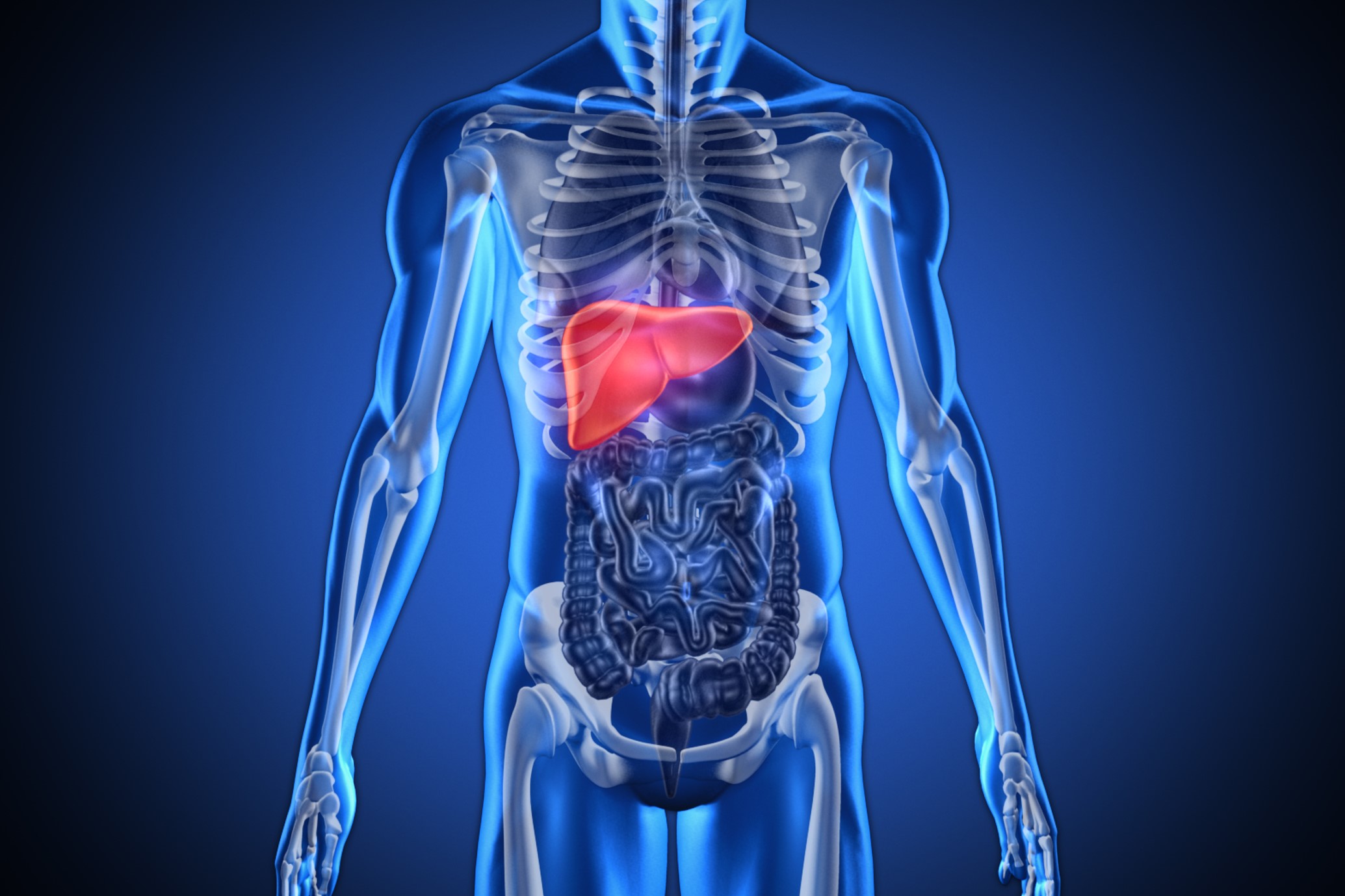
What it is and what are the symptoms of Focal Nodular Hyperplasia
Focal Nodular Hyperplasia is a benign tumour of the liver of hepatocellular origin that occurs much more rarely than an angioma
Most cases are observed in female patients, aged between 20 and 40 years, i.e. in the fertile period.
In recent years the prevalence of Focal Nodular Hyperplasia seems to have increased greatly
The ratio with the other benign hepatocellular tumour, liver adenoma, is now about 10 to 1: this epidemiological fact must be taken into account in the differential diagnosis between the two lesions.
Symptoms of benign liver tumour
Nodular hyperplasia remains asymptomatic in the vast majority of cases.
Symptoms, when present, are minor and non-specific, such as a sense of heaviness or vague postprandial abdominal pain.
The evolution of focal nodular hyperplasia is absolutely benign.
Rarely it may undergo a slow and progressive increase in volume.
Risks of spontaneous bleeding or malignant neoplastic transformation are absent.
Laboratory investigations are usually normal, except for rare cases in which a moderate increase in gamma-glutamyl-transpeptidase (Gamma GT) is present.
Diagnosis of focal nodular hyperplasia
Hepatic ultrasound remains the first-level investigation, but does not add any particular diagnostic elements to the discovery of the lesion.
Echo-Doppler study, on the other hand, can be decisive for the diagnosis.
Computed tomography (CT) can show very characteristic features.
Hepatic scintigraphy with technetium may be useful in the differential diagnosis with adenoma.
Nuclear magnetic resonance imaging (NMR) is the test with the greatest sensitivity and specificity in the diagnosis of nodular hyperplasia, even though the appearance of the lesion may be very varied.
On the usefulness of percutaneous biopsy for preoperative diagnosis of certainty, similar considerations to those already made for angioma apply.
Treatment of benign liver tumours
The same considerations made above for angiomas apply to nodular hyperplasia.
Surgical indications are limited to symptomatic forms, which account for about 15% of observed cases.
On the other hand, indications for diagnostic laparotomy appear less and less justified.
When the lesion has clinical features (young woman, without chronic hepatopathy, with negative neoplastic and viral markers) and radiological features (central scar with wagon wheel image and with septa separating homogeneous nodules), simple observation with periodic ultrasound checks may be sufficient.
This cautious attitude is advisable until one is absolutely certain that other neoplastic forms can be excluded.
Read Also:
Emergency Live Even More…Live: Download The New Free App Of Your Newspaper For IOS And Android
Complications Of Liver Cirrhosis: What Are They?
Neonatal Hepatitis: Symptoms, Diagnosis And Treatment
Cerebral Intoxications: Hepatic Or Porto-Systemic Encephalopathy
What Is Hashimoto’s Encephalopathy?
Bilirubin Encephalopathy (Kernicterus): Neonatal Jaundice With Bilirubin Infiltration Of The Brain
Hepatitis A: What It Is And How It Is Transmitted
Hepatitis B: Symptoms And Treatment
Hepatitis C: Causes, Symptoms And Treatment
Hepatitis D (Delta): Symptoms, Diagnosis, Treatment
Hepatitis E: What It Is And How Infection Occurs
Hepatitis In Children, Here Is What The Italian National Institute Of Health Says
Acute Hepatitis In Children, Maggiore (Bambino Gesù): ‘Jaundice A Wake-Up Call’
Nobel Prize For Medicine To Scientists Who Discovered Hepatitis C Virus
Hepatic Steatosis: What It Is And How To Prevent It
Acute Hepatitis And Kidney Injury Due To Energy Drink Consuption: Case Report
The Different Types Of Hepatitis: Prevention And Treatment
Hepatitis C: Causes, Symptoms And Treatment
Liver Cirrhosis: Causes And Symptoms
Hepatic Distomatosis: Transmission And Manifestation Of This Parasitosis


